Real case: necrotising fasciitis in a dog's thigh, managed with NPWT
Content note. The video shows real clinical wound photos from a clinician-reported case, shared with permission. They appear after a warning screen. The written guide below has no photos.
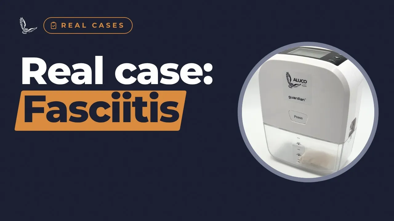
A clinician-reported case from Aura Veterinary: a dog with necrotising fasciitis of the inner thigh. After surgery to remove dead tissue, NPWT ran continuously at 125 mmHg for three days over an antimicrobial silver dressing, and the wound was then ready to plan a skin-flap closure. Contains a clinical photo. One case, not proof of effect.
Three things to remember
- A severe, deep infection, cleaned out in surgery.
- Three days of continuous NPWT over a silver dressing.
- Then a cleaner wound, ready to plan a skin-flap closure.
The full episode, written out
This is the narration from the video, so you can read it at your own pace, share it, or come back to a section.
Introduction
Welcome to Aluco Health. I'm Georgie Hollis. This is Real Cases, where we look at one clinician-reported case at a time. Today, a dog with necrotising fasciitis in the thigh, a severe infection of the deeper tissues.
The patient
This dog was referred to Aura Veterinary with necrotising fasciitis of the inner thigh. That's a severe infection of the deeper tissues that kills tissue as it spreads. The dog had bacteria in the bloodstream, severe swelling of the leg, patches of dead skin, and a lot of tissue coming away. The case was reported by Professor Nick Bacon, a specialist surgeon at Aura.
The challenge
The challenges were serious. The infection was deep, with dead tissue spreading quickly. The wound produced so much fluid that standard dressings were hard to keep in place. There was a lot of swelling and contamination. The team needed to remove dead tissue aggressively, without sedating the dog again and again. And the wound would later need closing with a skin flap.
The treatment
Under general anaesthetic, the wound was cleaned out extensively, removing dead tissue, and flushed. Then the team placed an antimicrobial silver dressing directly onto the wound bed. Aluco foam went over that layer, sealed with film, which held despite the wound's irregular shape. A suction port connected the dressing to the Aluco Guardian.
The settings
Suction was continuous, at one hundred and twenty-five millimetres of mercury, which the team describes as the standard pressure for contaminated or infected wounds. Therapy ran for three days without interruption. The seal was checked every day, and only needed minor reinforcement. Throughout, the dog stayed comfortable and stable.
After 72 hours
After seventy-two hours, the swelling had markedly reduced. The edges of the dead tissue were clearly marked out, so the team could see what was healthy. A healthy bed of granulation tissue was forming, and the wound looked visibly cleaner. The tissue had improved enough for the surgeons to plan a skin-flap reconstruction, and at the time of the report, the dog was booked in for that surgery.
Take-aways
What can vet teams take from this case? Here, an antimicrobial silver dressing sat under the foam, with NPWT working over the top. Check the seal every day. And hang the unit upright; the team notes this avoids false full-canister alarms. As the surgeon put it, for necrotising fasciitis they need aggressive fluid control and a reliable seal. Remember, this is one clinician-reported case. It shows what happened for this dog, not how other wounds will respond.
Words explained
- Necrotising fasciitis
- A severe infection of the deeper tissues that kills tissue as it spreads.
- Debridement
- Removing dead or contaminated tissue from a wound.
- Demarcated
- Clearly marked out, so dead and healthy tissue can be told apart.
- Skin flap
- Nearby skin moved over a wound while still attached at one edge.
- Granulation tissue
- New, healthy healing tissue (red and bumpy).
Sources
- S1Aluco Health. Case study 005: Necrotising fasciitis. Case provided by Nick Bacon, Aura Veterinary. Approved; published on alucohealth.com.
General information about animal wound care, not veterinary advice. Always follow your vet's advice. Aluco Guardian™ is for use by veterinary professionals. Evidence mentioned is not specific to Aluco Guardian™ unless we say so.
See the Aluco Guardian™ in your practice
Hire one for a single case, try it with a demonstration, or buy for a regular caseload. Training and round-the-clock clinical support for vet teams come with it.
Ask your vet whether NPWT could help
Your vet knows your pet best. Take your questions to them, or send them this page.
Watch next
 3:23
1 Dec
 1:59
1 Dec
 2:31
1 Dec
Aluco Guardian NPWT: how to change the canister
 4:40
1 Dec